
News|Articles|February 11, 2025
Lactate Is a Key Regulator of Immune Escape, Bone Marrow Fibrosis in Myelofibrosis
Author(s)Luke Halpern, Associate Editor
Listen
0:00 / 0:00
Key Takeaways
- Circulating lactate and MCT1 expression are key in immune escape and fibrotic transformation in myelofibrosis patients.
- Blocking MCT1 may serve as a novel antifibrotic strategy in myelofibrosis treatment.
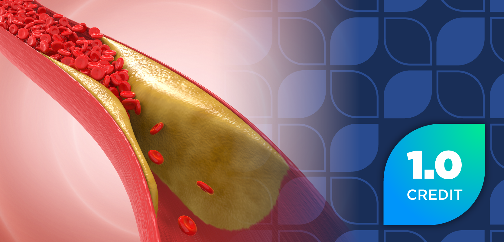
Circulating lactate is increased in patients with myelofibrosis and corresponds with the remodeling of lactate export channel monocarboxylate transporter 4, suggesting a link with fibrosis establishment.
Advertisement
New study results published in the Journal of Translational Medicine reveal that circulating lactate is a key regulator of immune escape and bone marrow (BM) fibrotic transformation in patients with myelofibrosis (MF), with a marked increase in lactate concentration and lactate import channel monocarboxylate transporter 1 (MCT1) expression in the site of blood cell production. The investigators suggest that MCT1 blocking could be used as a novel antifibrotic strategy in patients with MF.1
According to the study authors, the pathophysiology of MF is significantly influenced by alterations in the BM microenvironment, and these changes are often associated with the metabolic reprogramming of cancer cells in patients. Glycolysis is a hallmark aspect of the changes that occur in cancer cells; prior evidence from investigational models strongly suggests that lactate production is associated with tumor microenvironment (TME) progression in patients with certain types of cancer.1-3
Prior observations also indicate that increases in lactate dehydrogenase A (LDHA) have a central role in glycolysis, with high LDHA levels typically found in the sera of patients with MF, predicting shorter overall and leukemia-free survival. Within this context, excess lactate that is secreted by cancer cells can promote immune suppression and angiogenesis while also possibly playing a role in increasing the number of cancer-affiliated phenotypes (CAF), which promote fibrotic tumor formation, the investigators wrote.1,4
These authors sought to fill a gap in knowledge within the characterization of TME metabolic composition in patients with MF. Through their analysis, they demonstrated that the amount of circulating lactate increases in patients with MF, which eventually leads to the expansion of immunosuppressive subsets and the development of fibrosis. Furthermore, they observed that the expression of lactate export channel monocarboxylate transporter 4 (MCT4) in the TME becomes deeply remodeled during fibrotic transformation, “suggesting a link between lactate trafficking and pro-fibrotic establishment.”1
Upon measuring lactate concentration in patients with MF (n = 23) and healthy matched controls (HC; n = 16), the investigators found that the amount of circulating lactate was significantly increased in sera in patients with MF compared with HC (3.69 ± 2.2 vs 1.08 ± 0.3 mmol/L, P < 0.0001). Furthermore, after 48 hours of lactate exposure, there was a significant upregulation of alpha-1 type I collagen (Col1A1; P < 0.001 vs untreated cells), which favors the fibrotic transformation of the TME.1
Next, the investigators aimed to validate the downregulation of MCT4 in patients with MF by analyzing MCT expression in BM biopsies from patients with primary MF (PMF). Mesenchymal stromal cells (MSCs) play a major role in releasing profibrotic cytokines in patients with MF; therefore, they were scrutinized in the provided BM biopsies. The biopsies and subsequent analysis revealed that PMF-MSCs, which were identified as CD90+ cells, expressed only MCT1, indicating that the loss of MCT4 is an exclusive feature of the MF TME, according to the study authors.1
The data garnered from this trial are consistent with previous models indicating lactate’s role in promoting tumor progression in MF. Through the deep modulation of immune responses through immunosuppressive subset expansion, lactate prompts naïve T cell apoptosis and decreases cytotoxic activity, according to the investigators. However, the pathophysiology of underlying BM fibrosis in MF, despite intensive research, remains unclear.1-3
Of particular interest is the role of MCT4 as a diagnostic marker of MF, with the investigators noting the absence of MCT4 in MF, which is a unique aspect among compared with other myeloproliferative neoplasms. The authors discuss how MCT4 mediates lactate export in highly glycolytic cells, which could occur in patients with MF and support tumor progression. More research is required to further elucidate the role of MCT4 as a potential marker of clinical utility in MF diagnosis.1
REFERENCES
1. Spampinato M, Giallongo C, Giallongo S, et al. Lactate accumulation promotes immunosuppression and fibrotic transformation of bone marrow microenvironment in myelofibrosis. J Transl Med. 2025;23(69). doi:10.1186/s12967-025-06083-4
2. Sharma D, Singh M, Rani R. Role of LDH in tumor glycolysis: Regulation of LDHA by small molecules for cancer therapeutics. Seminars in Cancer Biology. 2022;87:184-195. doi:10.1016/j.semcancer.2022.11.007
3. Longhitano L, Vicario N, Tibullo D, et al. Lactate induces the expressions of MCT1 and HCAR1 to promote tumor growth and progression in glioblastoma. Front Oncol. 2022;12. doi:10.3389/fonc.2022.871798
4. Shah S, Mudireddy M, Hanson CA, et al. Marked elevation of serum lactate dehydrogenase in primary myelofibrosis: Clinical and prognostic correlates. Blood Can Journ. 2017;7:657. doi:10.1038/s41408-017-0024-9
Newsletter
Stay informed on drug updates, treatment guidelines, and pharmacy practice trends—subscribe to Pharmacy Times for weekly clinical insights.
Advertisement
Related Content
Advertisement


Latest CME


Advertisement
Advertisement
Trending on Pharmacy Times
1
TrumpRx Launches, Offering Cash-Paying Patients Discounted Drugs
2
Nipah Outbreak in India Poses Low Global Risk Despite Lack of Approved Treatments
3
Pharmacist Takeover: CAR T Therapy Signals a Shift in ALL Treatment
4
SGLT2 Inhibitors in T2D Lower 5-Year Risk of CKD and Acute Kidney Injury
5